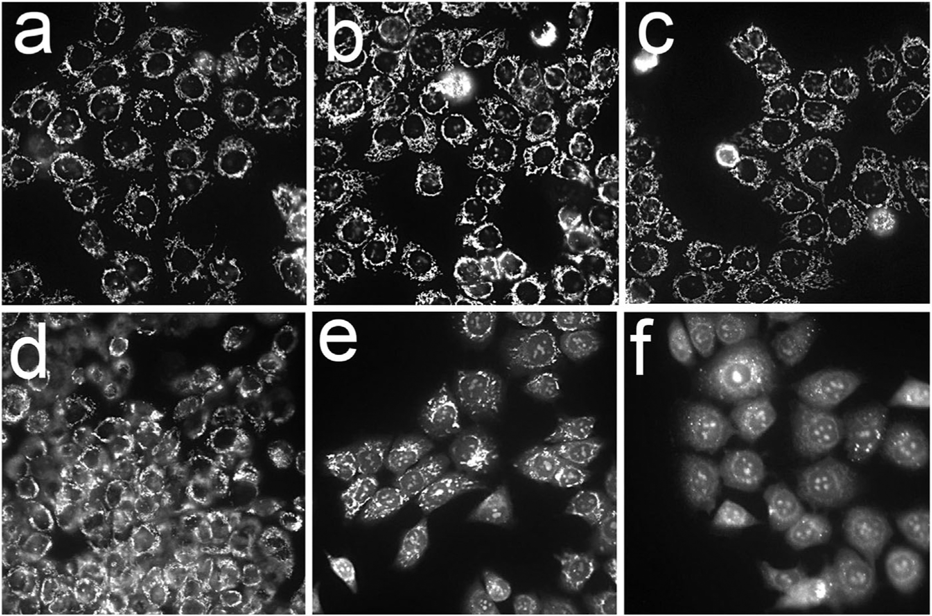
Fig. 4.

Photodynamic effects of different BPD formulations on the mitochondrial membrane potential. After 3 hours loading incubations, OVCAR5 cells were irradiated at 690 nm (200 mJ/ sq cm) with the MTO fluorescence labeling pattern determined directly after irradiation. a = untreated cells, b –BPDf; c, BPDa+c; d, BPDa−c; e, BPDf/BPDa+c; f, BPDf/BPDa-c. Extracellular levels of the photosensitizers are indicated in the legend to Figure 2.
