
Fig. 6.
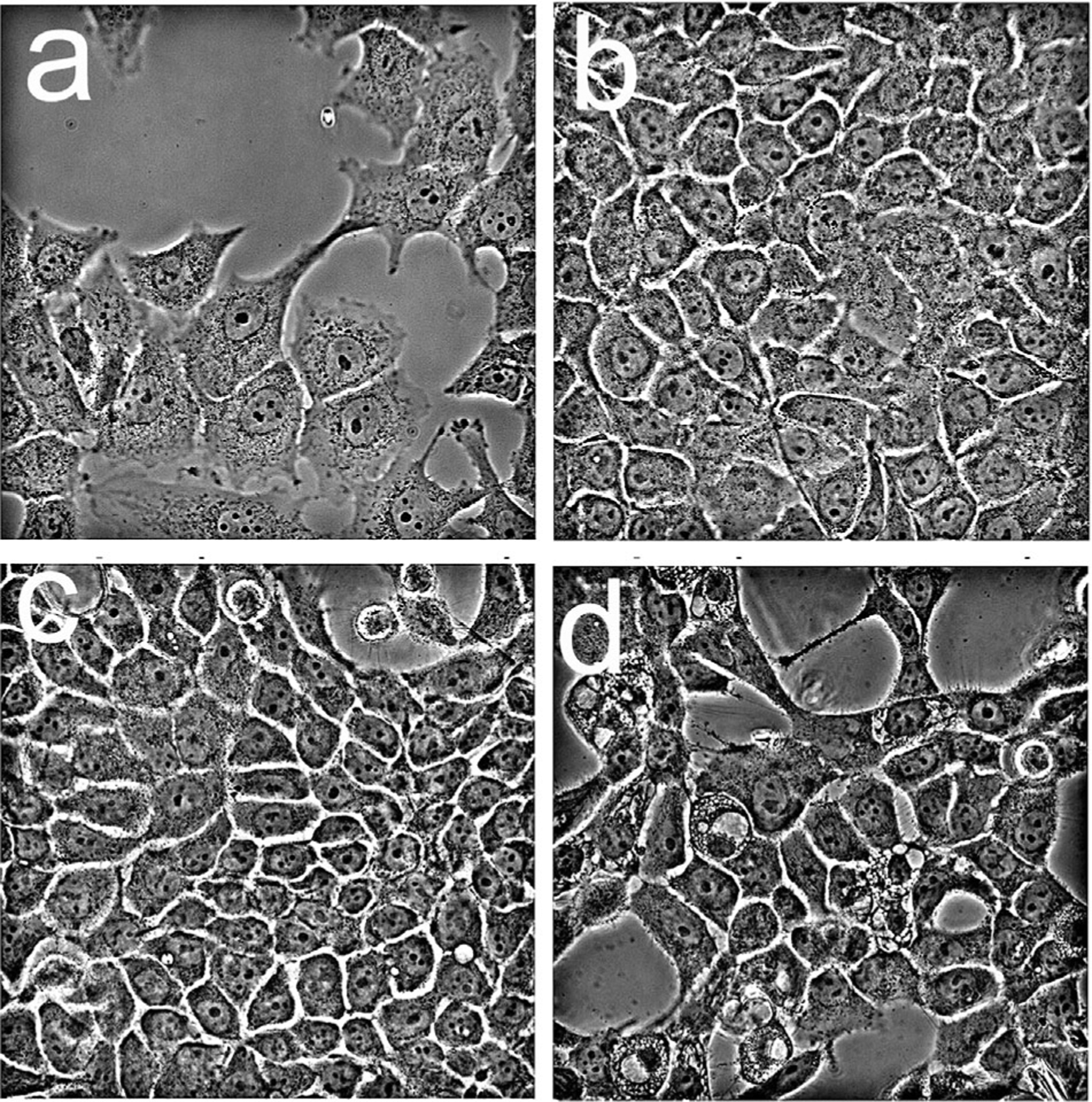
Morphology of OVCAR5 cells one day after irradiation as detected by phase-contrast microscopy. Cells were incubated with liposomal formulations of lipo-BPD (0.05 μM) and anchored formulations (0.5 μM) prior to irradiation (200 mJ/sq cm): a, untreated cells; b, BPDf; c, BPDf/BPDa+c; d, BPDf/BPDa−c.