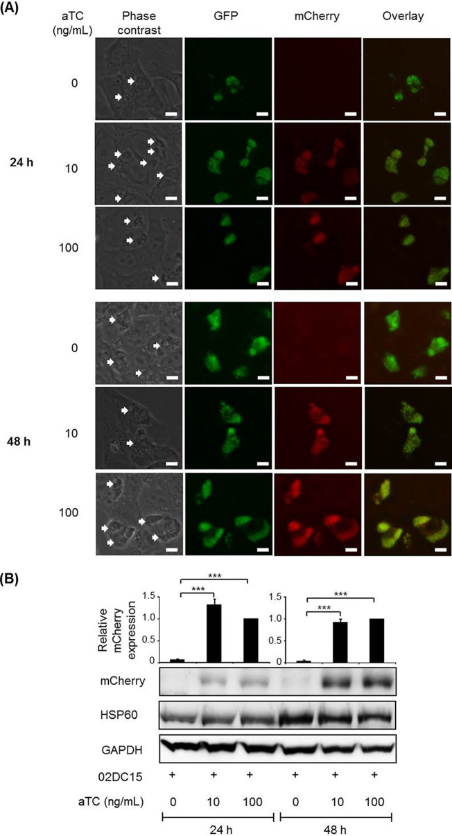
FIG 2.
GFP expression and mCherry induction in pCps-Tet-mCherry-transformed C. psittaci strain 02DC15. Transformed C. psittaci 02DC15 bacteria were grown in epithelial cells with 1U/ml PEN for 24 and 48 h. (A) GFP fluorescence of chlamydial inclusions was visualized in living cells without fixing and staining. aTC (10 and 100 ng/ml) was added at 1 hpi to induce mCherry expression. Images are representative of three independent experiments. White arrows show chlamydial inclusions. White scale bars represent 10 μm. (B) mCherry was analyzed by Western blotting and densitometric analyses at 24 and 48 hpi. mCherry protein amounts were normalized to chlamydial HSP60. GAPDH was used as a loading control. (n = 4; mean ± SEM; Sidak’s multiple comparison: ***, P ≤ 0.001).

