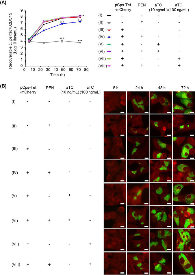
FIG 3.
One-step growth curve and inclusion morphology of pCps-Tet-mCherry-transformed and untransformed C. psittaci strain 02DC15. (A) C. psittaci strain 02DC15 transformed or untransformed with pCps-Tet-mCherry was grown in epithelial cells with or without 10 U/ml PEN and 0, 10, or 100 ng/ml of aTC. Recoverable C. psittaci 02DC15 IFUs were determined at 5, 24, 48, and 72 hpi. The numbers of recoverable C. psittaci 02DC15 under each condition (II to VIII) at the indicated time were compared to those of untransformed C. psittaci 02DC15 without PEN (I). (n = 3 to 6; mean ± SEM; Sidak’s multiple comparison: **, P ≤ 0.01; ***, P ≤ 0.001). (B) Representative immunofluorescence images of pCps-Tet-mCherry-transformed or untransformed C. psittaci 02DC15 at 5, 24, 48, and 72 hpi. Chlamydial inclusions were stained by FITC-labeled monoclonal chlamydial-LPS antibodies. Evans blue counterstaining of host cells was used for better characterization of intracellular inclusions. Images are representative of 3 to 6 independent experiments. White scale bars represent 10 μm.

