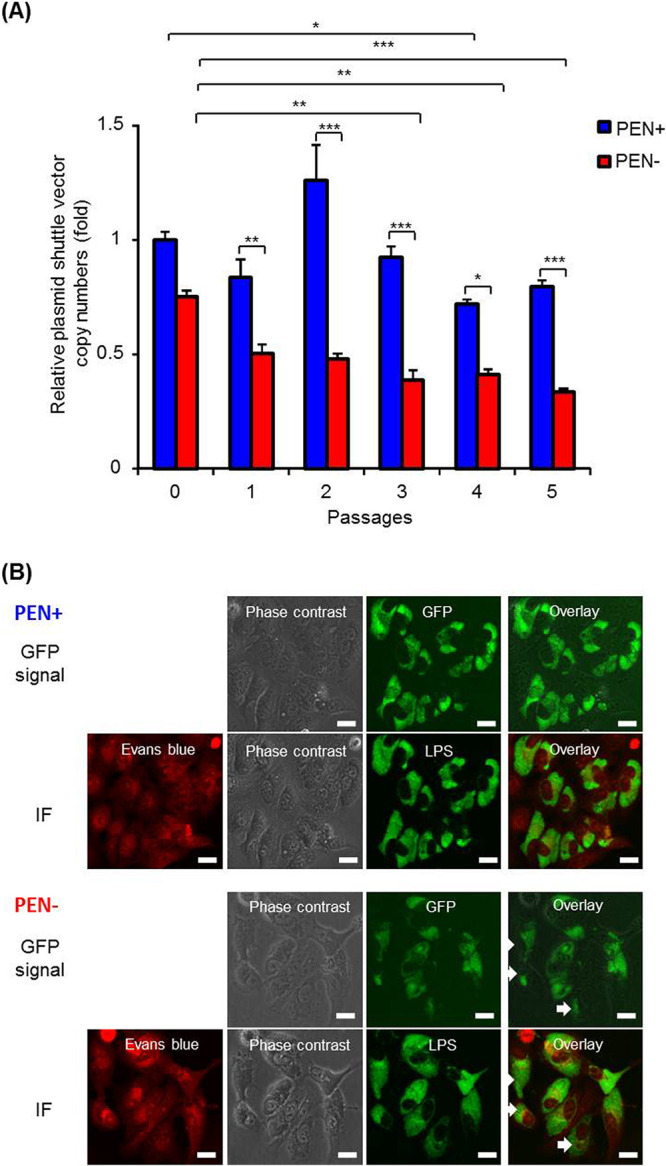
FIG 4.

The pCps-Tet-mCherry plasmid can be stably maintained by C. psittaci strain 02DC15 and expresses GFP. (A) pCps-Tet-mCherry-transformed C. psittaci 02DC15 bacteria were subcultured in epithelial cells in the presence or absence of 10 U/ml PEN every 2 days over 5 passages. Quantitative PCR was performed using primers specific to genomic DNA or to the pCps-Tet-mCherry plasmid. Copy numbers of pCps-Tet-mCherry were normalized to genomic DNA at each passage in the presence or absence of PEN, and were compared to passage 0 of C. psittaci 02DC15 in the presence of PEN. (n = 3; mean ± SEM, Sidak’s multiple comparison: *, P ≤ 0.05; **, P ≤ 0.01; ***, P ≤ 0.001). (B) Representative GFP and immunofluorescence images of pCps-Tet-mCherry-transformed C. psittaci 02DC15 cells 48 hpi at passage 5. After a GFP signal was detected by fluorescence microscopy, the cells were fixed by methanol. Then, chlamydial inclusions were stained by FITC-labeled monoclonal chlamydial-LPS antibodies. Evans blue counterstaining of host cells was used for better characterization of intracellular inclusions. IF, immunofluorescence; LPS, lipopolysaccharide. White arrows show C. psittaci 02DC15 that lost pCps-Tet-mCherry during passages. White scale bars represent 20 μm.
