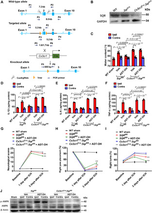
Fig. 6. SQR mediates therapeutic effects of H2S via AMPK activation following ICH in vivo.

(A) Schema illustrating generation of mice with microglia/macrophage-specific deletion of SQR (Cx3cr1cre:Sqrfl/fl). (B) Immunoblot analysis of SQR expression on primary microglia from Cx3cr1cre:Sqrfl/fl mice, floxed control mice (Sqrfl/fl), and wild-type mice (WT); three independent replicates. (C) Brain edema assessed by measuring water content in the ipsilateral (hemorrhagic) striatum (Ipsil) and contralateral striatum (Contra) at 3 days following ICH (n = 6). WT sham, sham-operated wild-type mice. ADT-OH (50 mg/kg per day) was administered for 3 days, starting at 3 hours after ICH. (D to F) Protein levels of interleukin-1β (IL-1β), IL-6, and tumor necrosis factor–α (TNF-α) in the striatum 3 days following ICH (n = 4). (G to I) Neurological deficits assessed by the neurological score, forelimb placement, and corner test following ICH (n = 6). **P < 0.01, indicating a statistically significant difference between the control mice treated with Veh (Sqrfl/fl) and the control mice treated with ADT-OH (Sqrfl/fl + ADT-OH) at 3 days after ICH. No statistical difference was detected between Veh-treated Cx3cr1cre:Sqrfl/fl mice (Cx3cr1cre:Sqrfl/fl) and ADT-OH–treated Cx3cr1cre:Sqrfl/fl mice (Cx3cr1cre:Sqrfl/fl + ADT-OH). (J) AMPK activation in the striatum of the mice treated with ADT-OH or Veh at 3 days following ICH (three independent replicates).
