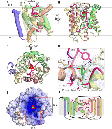
Fig. 2. Crystal structure of YeeE.

(A and B) Overall structure of StYeeE in cartoon (A) and ribbon (B) representations from the membrane side. The transparency of H1, H3, H8, and H10 in (A) is 30% to clearly display the inside. Numbers of α helices are indicated, and YeeE characteristic loops (LA–LD) are highlighted in red and magenta. Thiosulfate ions are shown as a space-filling model. (C) YeeE structure in ribbon representation from the outside. (D) Close-up view of thiosulfate-binding site. 2Fo − Fc map and thiosulfate-omit (Fo − Fc) map are shown with 1.2 σ and 4.0 σ, respectively. (E) Surface model of YeeE viewed from the outside, colored to indicate electrostatic potential ranging from blue (+10 kT/e) to red (−10 kT/e). (F) Schematic topology model of YeeE. YeeE has four short α helices (H2, H5, H9, and H12), the LA–LD loop, and nine transmembrane α helices (H1, H3, H4, H6, H7, H8, H10, H11, and H13).
