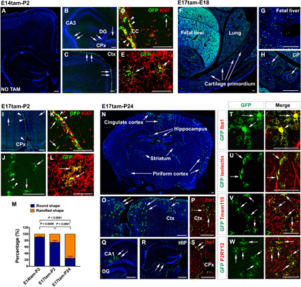
Fig. 4. The progeny of fetal CCR2+ monocytes in bitransgenic CCR2-CreER mice.

(A to E) Fate mapping CCR2+ monocytes in P2 mouse brains after tamoxifen dosing at E14 (n = 5). No GFP+ cells were labeled without tamoxifen dosing (A; n = 4). With tamoxifen, GFP+ cells were present in the CPx, the subdural meninges, and the fornix (arrows in B and C). GFP+ monocyte derivatives include Ki67+ cells in subdural meninges (arrowhead in D) and ramified microglial cells (arrows in D and E). The ramified monocyte derivatives were P2RY12+ (arrows in E). (F to H) With tamoxifen at E17, GFP+ cells were seen in the fetal liver and CP in E18 embryos (n = 6). (I to L) Fate mapping in P2 brains after tamoxifen induction at E17 (n = 5) showed GFP+ cells in the CPx and subdural meninges (arrowhead in I and J), and few ramified GFP+ cells (arrows in I to L). The progeny of CCR2+ monocytes included Ki67+ cells in subdural meninges (K) and P2RY12+ microglia (arrows in L). (M) Quantification of round- versus ramified-shaped progeny of fetal monocytes in E14tam-P2, E17tam-P2, and E17tam-P24 fate mapping (n = 5 for each). Data are shown as means ± SEM, one-way ANOVA with Tukey’s post hoc test. (N to W) Fate mapping monocyte derivatives in P24 brains after tamoxifen dosing at E17 showed Iba1+GFP+ microglia in the cingulate cortex, piriform cortex, striatum, and the hippocampus (N to Q and T; n = 5). Meningeal macrophages (R and S) and perivascular microglia (U) were visible. GFP+ ramified monocyte derivatives were Tmem119+ (V) and P2RY12+ (W). Scale bars, 200 μm (F to H), 100 μm (A to E, (I to L, and N to W).
