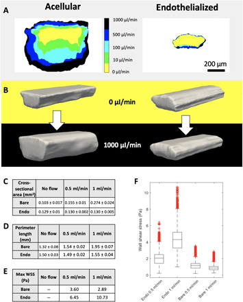
Fig. 2. Endothelial cells can remodel bioprinted vascular beds.

(A) Cross-sectional area collected at increasing flow rates for both acellular and endothelialized printed vessels. (B) Rendered 3D reconstructions of confocal image stacks of both acellular and endothelialized devices under static conditions and exposed to flow (1 ml/min). (C) Vessel volume measurements collected from rendered 3D vessel reconstructions with SDs of 400 measurements at different positions along the vessel length. (D) Vessel perimeter length measurements collected from rendered 3D vessel reconstructions with SDs of 400 measurements along the vessel length. (E) Maximum WSS values calculated with HARVEY from rendered vessels at different flow rates show that the deformable acellular vessel experiences lower WSS with higher flow, whereas the relatively stiff endothelialized vessel experiences fivefold greater peak WSS with 2× increase in flow. (F) WSS distribution in endothelialized geometry increases with increased flow, whereas lower WSS is observed in the acellular geometry.
