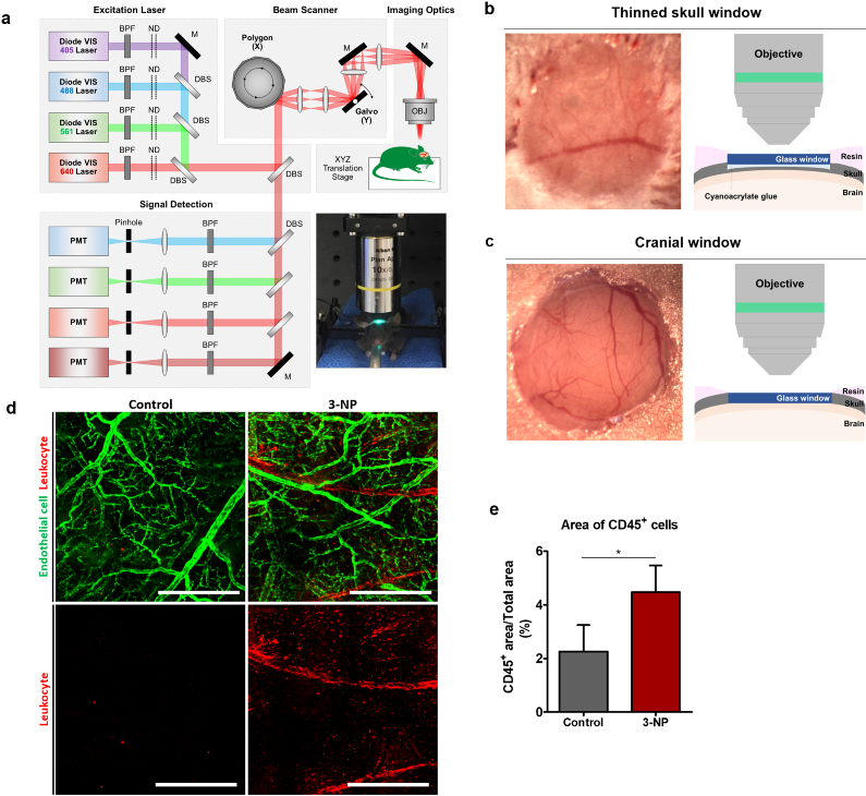
Fig. 1.
(a) Schematic of custom-built video-rate laser-scanning confocal microscopy system (ND, neutral density filter; DBS, dichroic beam splitter; BPF, band pass filter; M, mirror; PMT, photomultiplier tube; OBJ, objective lens). (b-c) Photo and schematic of (b) thinned skull window and (c) chronic cranial window for intravital imaging of the brain. (d) Representative mosaic images obtained through the thinned skull of the Tie2-GFP mice in vivo; saline-injected control group and 3-NP injected group (green, Tie2-GFP expressed in vascular endothelial cells; red, leukocytes labelled by anti-CD45 antibody conjugated with Alexa 647). (e) Ratio of CD45+ cell occupied area over total imaged area in the control group (n = 4 field of views from 4 mice) and 3-NP group (n = 4 field of views from 4 mice). Scale bars = 500 μm.

