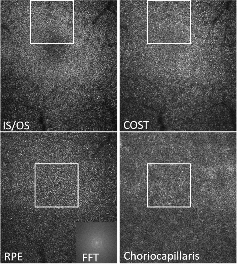
Fig. 3.
AO-OCT en-face images retrieved by depth integration of the layers shown in Fig. 2(b)). The white squares indicate region of interests that are presented as magnified views in Fig. 4. Field of view: 3.8° × 4°. IS/OS: junction between inner and outer segments of photoreceptors, COST: cone outer segment tips, RPE: retinal pigment epithelium, CC: choriocapillaris. FFT: 2D Fourier transform of the entire RPE image. In the lower row, the fovea centrals is located within the white square.

