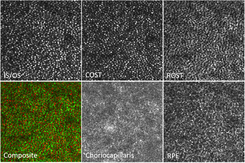
Fig. 8.
Magnified view of the regions of interest indicated with the white squares in Fig. 7. IS/OS: junction between inner and outer segments of photoreceptors, COST: cone outer segment tips, ROST: rod outer segment tips, Composite: false color image of COST (red) and ROST (green), RPE: retinal pigment epithelium, CC: choriocapillaris. The dimension of the magnified image is 1.3° × 1.3°.

