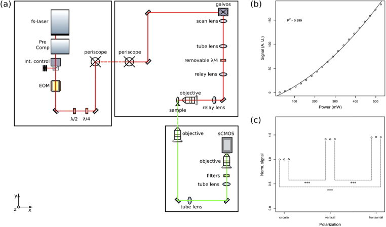
Fig. 2.
(a) Schematic of the custom-made 2P LS microscope. Fs-laser: femtosecond laser. Pre Comp: pulse compressor. Int. control: intensity control assembly, composed by a half-wave plate and a Glan–Thompson prism. EOM: Electro-Optical Modulator. λ/2: half-wave plate. λ/4: quarter-wave plate. Galvos: galvanometric mirror assembly, composed by a resonant mirror and a closed-loop mirror. Red line: excitation light. Green line: fluorescence light. The dashed lines indicate vertical paths. Axis specification is consistent with Fig. 1, with the excitation and detection objectives oriented along the x-axis and the z-axis, respectively. (b) Scatter plot of the fluorescent signal generated by a fixed Tg(actin:EGFP) larva as a function of the excitation power. Parabolic fit of the data is indicated by the continuous line and its coefficient of determination is reported on the graph. (c) Scatter plot of the signal generated by a fluorescein solution excited with circularly-, vertically- or horizontally-polarized light. Each condition was tested in triplicate and each point represents a single measure. The signal value is normalized to the average of the circular polarization case. Statistically significant differences are indicated by three asterisks.

