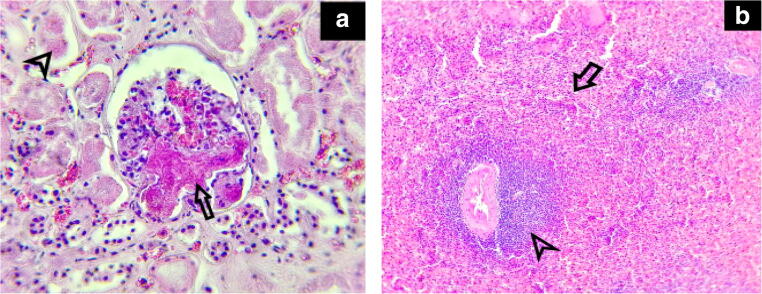
Fig. 2.
Microscopic aspect kidney and spleen (case 1)—hematoxylin-eosin (× 100, × 400). a Kidney (× 400) Focal microthrombi formation within the glomerular capillary lumen (arrow). Acute tubular injury (arrow head). b Spleen (× 100) Marked congestion with white pulp atrophy (arrow), the remaining lymphoid tissue was found around the central arteries (arrow head)

