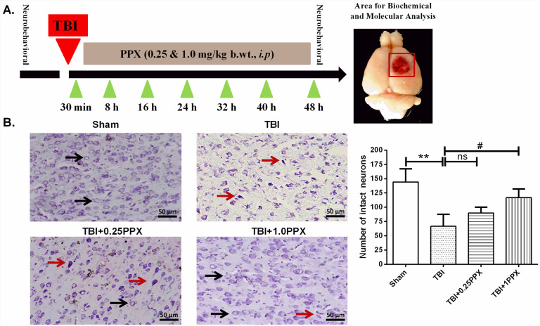
Fig. 1.
PPX administration reduces neuronal damage after TBI. (A) Timeline of the experimental treatment regimen used in this study (left). The area of the rat brain used for molecular and biochemical analysis is also shown (right). (B) Effects of PPX treatment on histological deficits in TBI rats. The arrows indicate the morphology of normal (black) and damaged (red) neurons in cortical rat brain tissue. In the sham group, neurons were intact with a clear border and a light stain. In the TBI group, the number of intact neurons was decreased, and the number of damaged neurons demonstrating cell shrinkage and dark staining was increased. PPX treatment increased the number of intact neurons. **P<0.01 compared with the sham group; #P<0.05 compared with the TBI group; ns, not significant (one-way ANOVA with Tukey's multiple comparisons test). Data are presented as mean±s.d. (n=3). Magnification at 20×.

