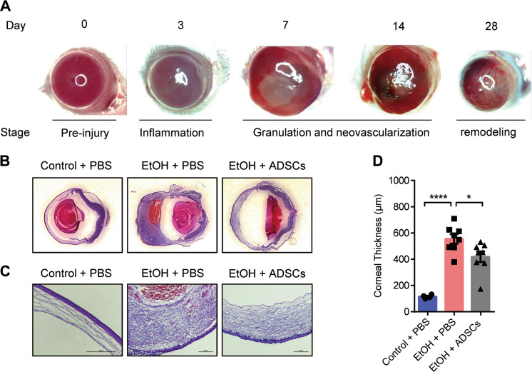
Fig. 2. ADSC treatment reduces corneal thickening during granulation.
a Representative images of the eyeball at different stages of corneal wound healing in the mouse CWH model. b, c Hematoxylin and eosin staining were performed. The images of whole eyeballs are shown in panel B, and the images of corneas are displayed in panel C (the magnification of the Control + PBS group is 20× and the other two groups are 10×, Scale bar, 200 μm). Mice (n = 4–8/group) were treated as described in Fig. 1. The thickness of corneas was measured (d). Data are shown as means ± SEM, *P < 0.05; ***P < 0.001 determined by one-way ANOVA with Tukey comparisons.

