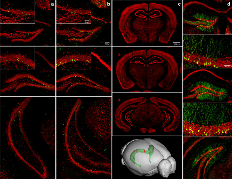
Fig. 1. Adult neurogenesis in the dentate gyrus of the hippocampus.
a, b Photomicrographs of brain sections throughout the rostral-caudal extent of the hippocampal dentate gyrus (DG), immunofluorescently double-labeled with BrdU (green) and NeuN (red), derived from adult C57Bl/6 mice housed under (a) control or (b) running conditions for 4 weeks. BrdU (50 mg/kg, i.p.) was injected daily for the first 10 days. The BrdU+ cell numbers in the DG granule cell layer are (a) 22, 46, 41, and (b) 44, 120, 52, respectively, from top to bottom. c Representative overview panels (distance from Bregma95 rostral to caudal: 1: −1.34 mm, 2: −1.94 mm, 3: −3.64 mm) of coronal sections through the hippocampus, labeled with NeuN (red). Lower panel: 3-D brain diagram shows the hippocampus (green) with numbers (1,2,3, red) corresponding approximately to section bregma levels in the panels above. d Photomicrographs of adult-born DG neurons with elaborate dendritic processes, targeted by retrovirus expressing green fluorescent protein (GFP) at one month after stereotaxic injection of retrovirus into the DG. Retrovirus labels dividing cells that develop into neurons over weeks. Coronal sections (40 μm), derived from adult C57Bl/6 mice, were co-labeled with NeuN (red).

