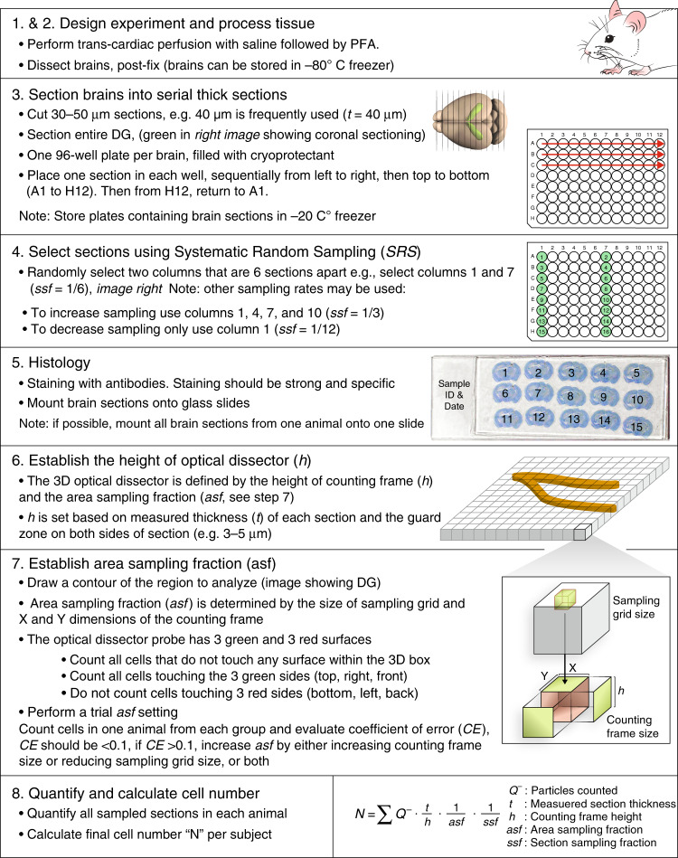
Fig. 3. Design-based stereology work flow for quantitative analysis of adult neurogenesis.
Optimal results of experiments can be obtained by following these steps: 1. Experimental design. 2. Tissue processing for optimal histological analysis. 3. Sectioning into thick (e.g., 40 μm) sequential sections and store in long-term tissue storage reagent (e.g. cryoprotectant). 4. Selection of representative sections using systematic random sampling (SRS). 5. Histological staining of the tissue with appropriate antibodies for cell types of interest. 6. Establishing the height of counting frame for optical fractionator (h). 7. Establishing the area sampling fraction (asf) to define the fraction of the specimen that will be quantified to represent the entire specimen. This is done by setting the sampling grid and defining the size of the counting frame (X and Y dimensions) for optical fractionator. 8. Quantification of sampled regions using optical fractionator and calculation of total numbers of markers in the specimen using the parameters set above and the number of cells counted.

