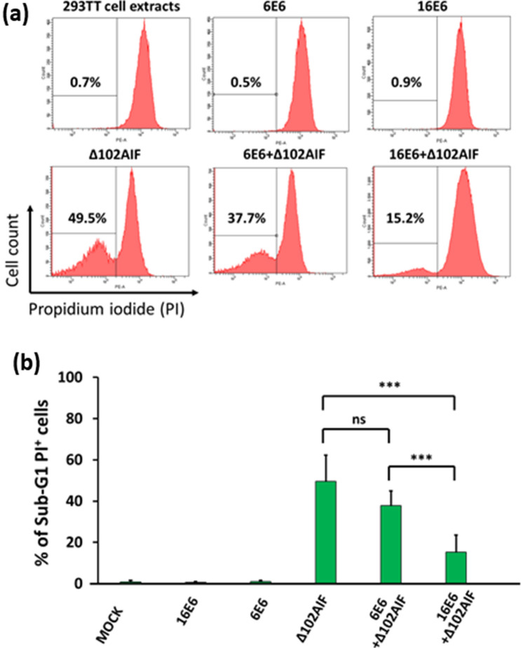
Figure 6.
Inhibition of AIF-induced chromatin degradation by HPV16 E6. 293TT cells were transfected with p16E6, p6E6, and/or pΔ102AIF expression plasmids. Then, cell lysates from the transfected cells were incubated with nuclei isolated from HeLa cells and stained with propidium iodide (PI). Chromatin degradation was assessed by flow cytometry and quantified by sub-G1 population gating. (a) Representative flow cytometric panels are shown. (b) The percentages of sub-G1 PI-positive cells from three independent experiments performed with five samples were analyzed, *** indicates a significant difference between the two groups. ns, no significant difference. Statistical analyses were performed by Krushal-Wallis test with Steel–Dwass test.

