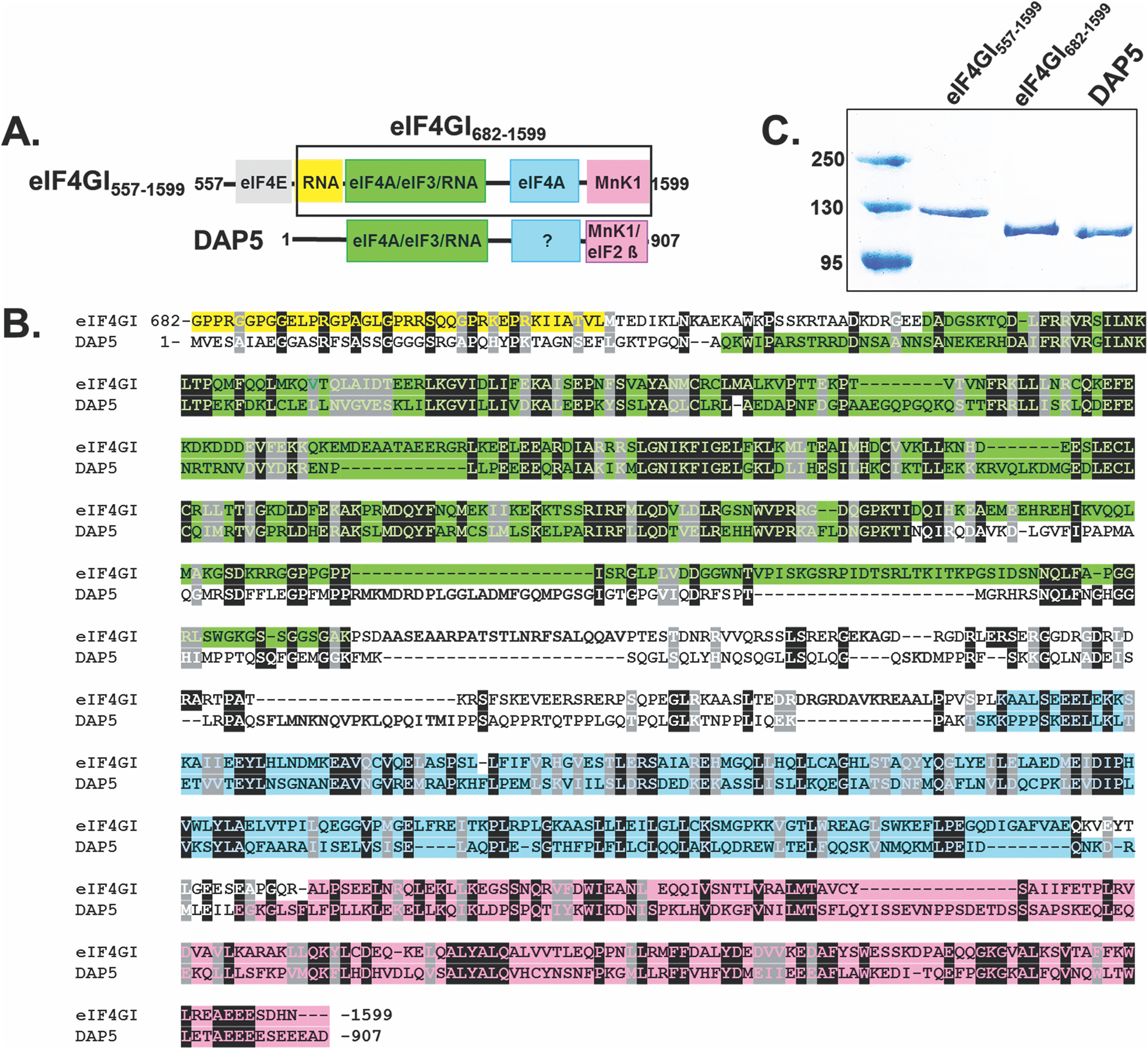
Figure 1.

Domain structure and sequence alignment between eIF4GI and DAP5. A, cartoons showing the domain architecture of eIF4GI557-1599 and full-length DAP5. eIF4GI contains a second eIF4A binding region in the MA3 domain (blue box in the eIF4GI cartoon), but the corresponding domain in DAP5 (blue box with question mark) does not bind to eIF4A and has unknown function (22). The shorter construct of eIF4GI, eIF4GI682-1599, is highlighted in the black box. B, sequence alignment encompassing similar domains in eIF4GI682-1599 and DAP5. The sequence alignment was performed using T-coffee (61). Residues are color-coded according to their domain organization shown in panel A. C, a 10% SDS-PAGE gel showing purity of eIF4GI557-1599, eIF4GI682-1599, and DAP5 used for this study.
