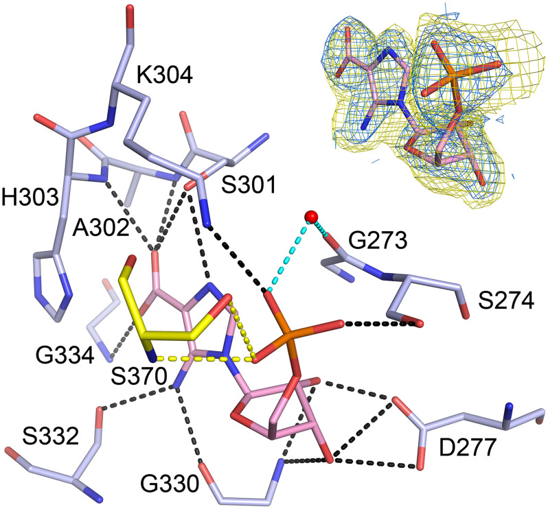
Figure 4.
CAIR bound in the AIRc active site. The CAIR molecule is shown as pink sticks and the active site residues involved in direct (black dashed lines) or water-mediated (cyan dashed lines) hydrogen bonds are shown as pale blue sticks. Residue Ser-370 from another PAICS monomer in the octamer that participates in ligand binding through hydrogen bonds (yellow dashed lines) is shown as yellow sticks. Refined 2Fo – Fc electron density map for CAIR contoured at 1σ and mFo – DFc Polder omit map for CAIR contoured at 4 σ are shown in the upper right corner in blue and yellow, respectively.

