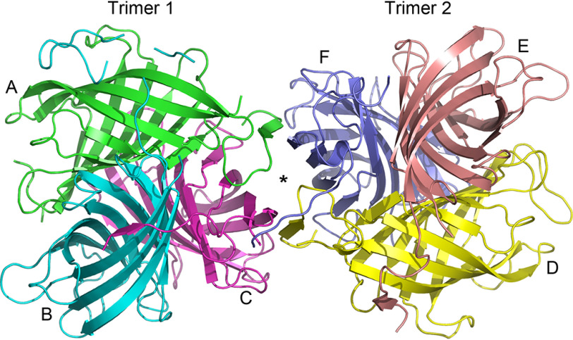
Figure 4.
Ribbon representation of the GePTS1 structure. The biological unit is a tightly packed trimer, and the asymmetric unit contains a dimer of trimers related by a pseudo-2-fold axis perpendicular to the plane of the paper. Trimer 1 on the left comprises monomers A (green), B (cyan), and C (magenta), and trimer 2 comprises monomers D (yellow), E (pink), and F (blue) on the right.

