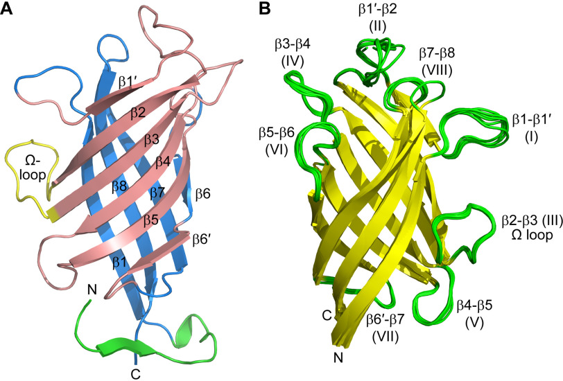
Figure 5.
Structure of the GePTS1 monomers. A, ribbon representation of the eight-stranded barrel colored as two β-sheets in pink and blue. The N-terminal region is colored green. An Ω loop between strands β2 and β3 is colored yellow. The secondary structure labeling is also shown. B, superposition of the six monomers onto each other.

