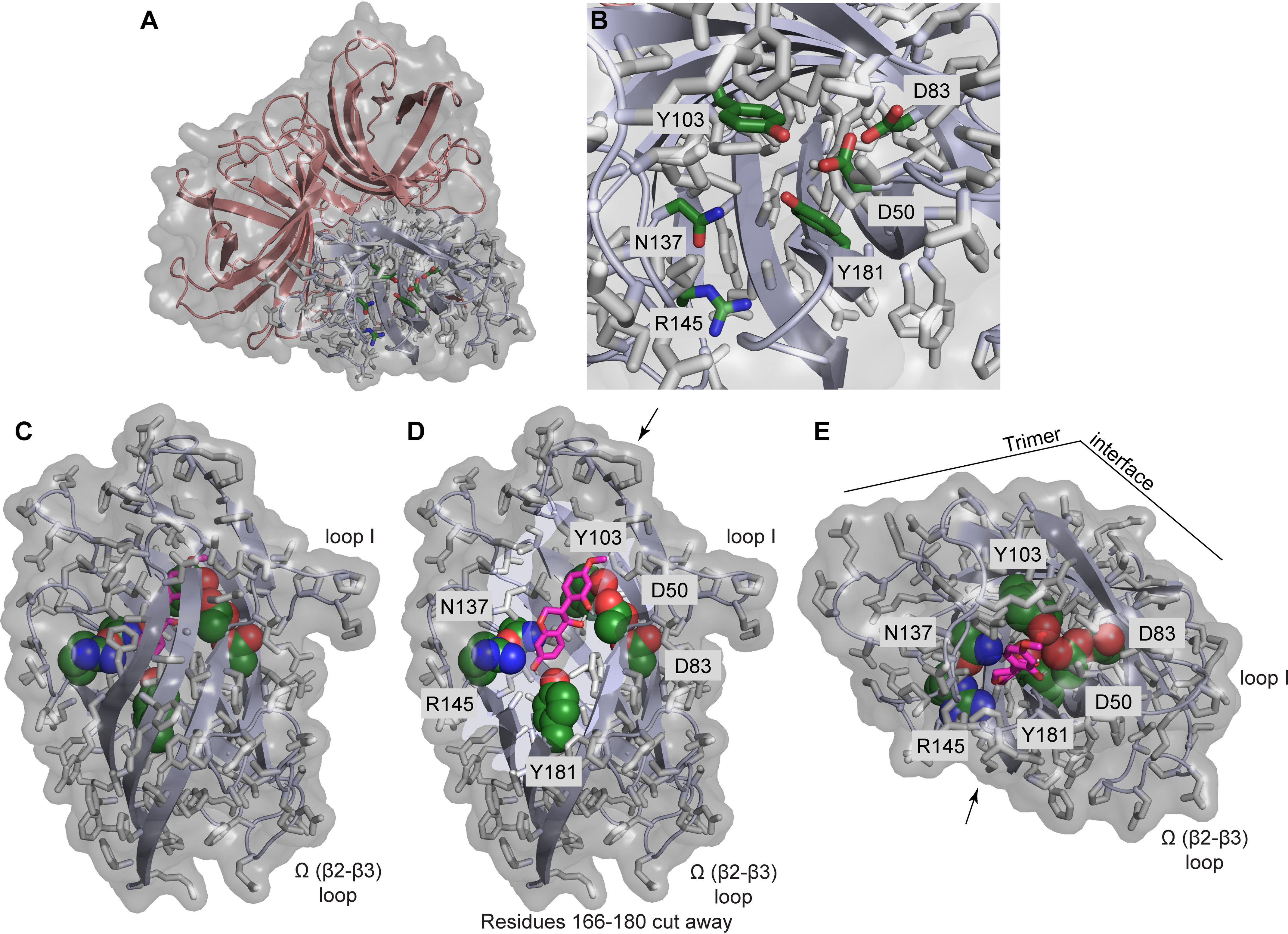
Figure 6.

The GePTS1 active site. A, depiction of the GePTS1 trimer in cartoon mode with transparent surface, viewed from the top near the 3-fold symmetry axis, showing side-chains of polar active-site residues (Asp50, Asp83, Tyr103, Asn137, Arg145, and Tyr181) for one monomer as sticks with dark green carbon atoms. B, zoom-in view of the active site. C, side view of GePTS1 monomer with docked (3S,4R)-DMI substrate (pink carbon atoms) indicating the degree to which the substrate can be buried within the barrel interior. D, same side view with residues 166–180 cut away to reveal the active-site tunnel with docked (3S,4R)-DMI and the polar active-site residues. E, top view of GePTS1 monomer with docked (3S,4R)-DMI, looking directly into the active-site tunnel. Arrows in D and E indicate the viewer's perspective shown in the other panel, and key loops and the trimer interface are indicated as reference points. The PsPTS1 active site is essentially identical to that of GePTS1 in terms of the residues present and their side-chain rotamers.
