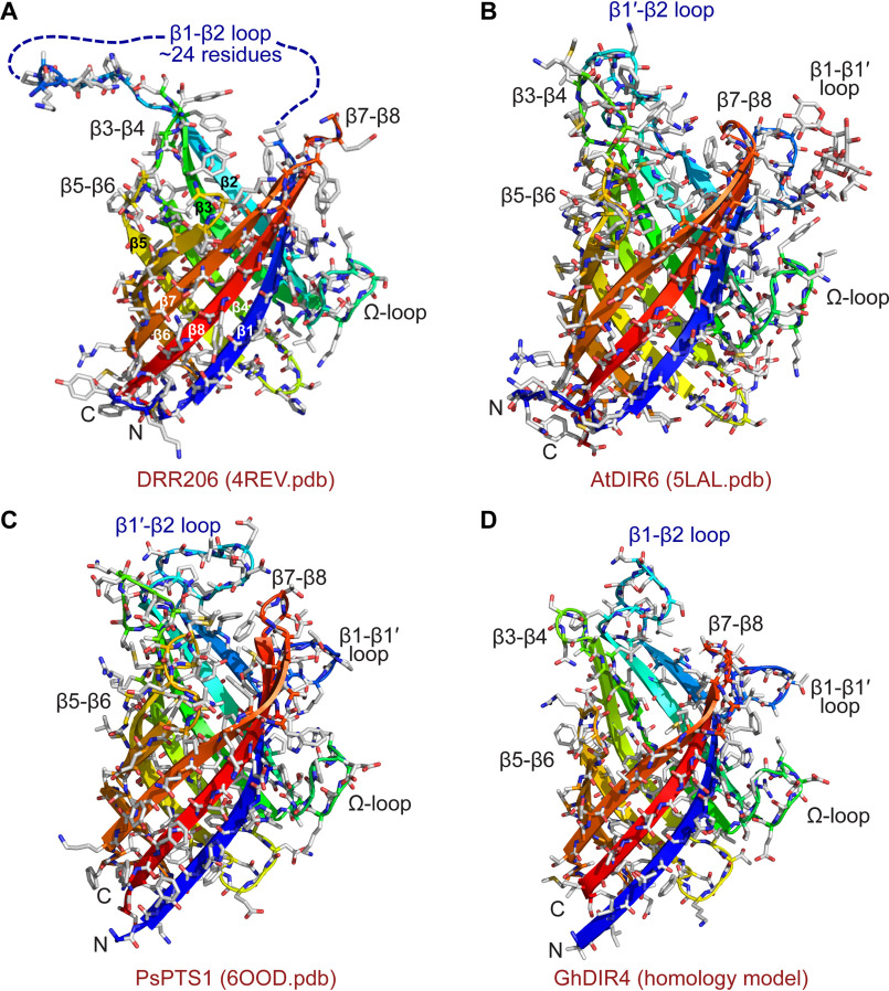
Figure 7.
3D structure and homology modeling of DPs. Shown are 3D structures of DRR206 (4REV) (A), AtDIR6 (5LAL) (B), and PsPTS1 (6OOD) (C). D, homology model of GhDIR4 created with Phyre2 in one-to-one threading mode using PsPTS1 structure as a template (30, 31). The β-strands are colored blue to red from the N to the C terminus: royal blue, β-1; slightly lighter blue, β1′; light blue-green, β2; green, β3; yellow-green, β4; yellow, β5; lighter orange, β6 and β6′; darker orange, β7; red, β8.

