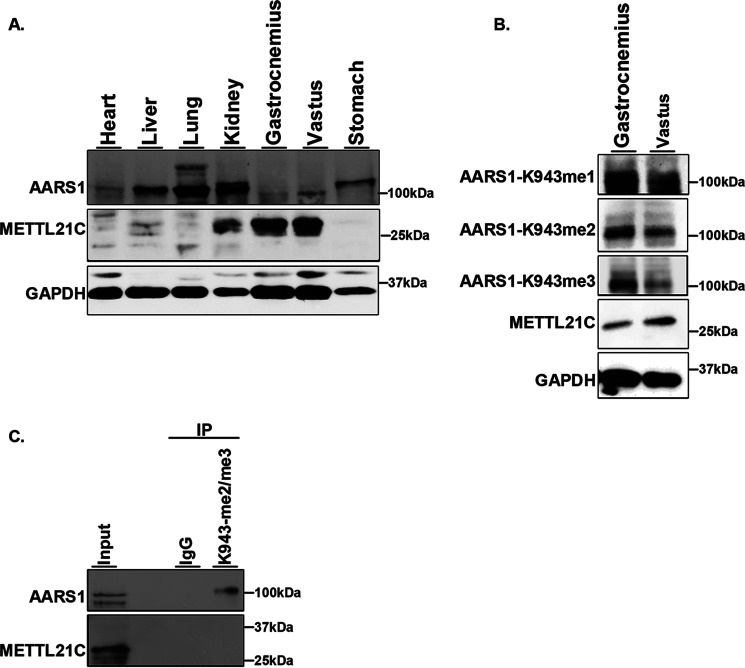
Figure 5.
AARS1 is methylated mouse muscle tissue. A, analysis of METTL21C and AARS1 protein levels in mouse tissues. Western blots of the indicated tissue with the indicated antibodies is shown. GAPDH is shown as a loading control. B and C, AARS1 is methylated in skeletal mouse tissue. B, Western blots of the indicated skeletal muscle tissues and the indicated antibodies. GAPDH is shown as a loading control. C, AARS1 is detected in IPs using the anti-AARS1-K943me2 and -me3 antibodies. Western blotting of the indicated IPs with the indicated antibodies is shown. IgG is used as negative control.

