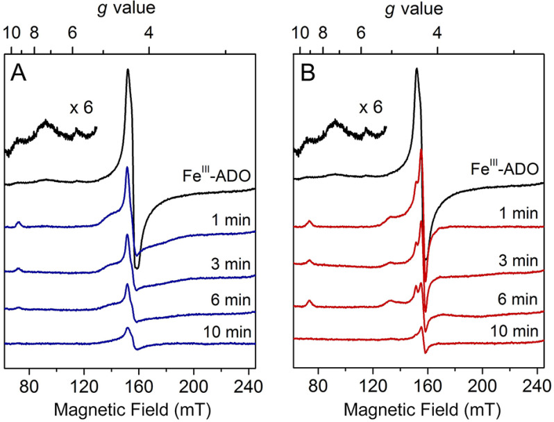
Figure 2.
Continuous-wave EPR spectra of time-resolved incubation of oxidized human ADO with cysteamine (A) and N terminus (B) of RGS5. Oxidized FeIII-ADO exhibited a major high-spin (S = 5/2) signal centered at g = 4.30 with multiple low-field resonances (inset, amplified 6 times) (black trace). FeIII-ADO was incubated with 30 mm cysteamine (blue traces) and RGS5 peptide (red traces) for 1, 3, 6, and 10 min. The EPR signal intensity is present in arbitrary units. Spectra were recorded at 10 K with 0.2 mW microwave power.

