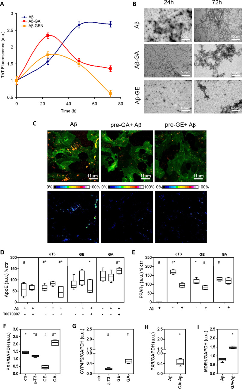
Figure 2.
Aβ aggregation and metabolism in mouse cortical astrocytes treated with genistein or garcinoic acid. A, ThT fluorescent test was utilized to assess cross-β-sheet structure of Aβ(1–42) during formation of amyloid aggregates in cell-free experiments. Fluorescence was investigated for 72 h in the absence or in the presence of GE (5 μm) or GA (25 μm). B, Structural aspects of Aβ aggregation were investigated by transmission EM; scale bars, 500 μm. Aβ(1–42) aggregates on the plasma membrane of mouse astrocytes pre-treated with test molecules were assessed by immunofluorescence. Scale bars, 11 μm. D–I, immunoblot of extracellular ApoE and cellular levels of PPARγ, PXR, CYP4F2, and MDR1. Determinations were carried out in mouse cortical astrocytes pre-treated with GE (5 μm), GA (25 μm) or δ-T3 (2.5 μm), and then exposed to Aβ. In some experiments, the effect of the PPARγ activity inhibitor T0070907 was also investigated (D). Further details on cell treatments and determinations are reported under :Experimental procedures.” #, p < 0.05 versus Ctr test; *, p < 0.05 versus Aβ test.

