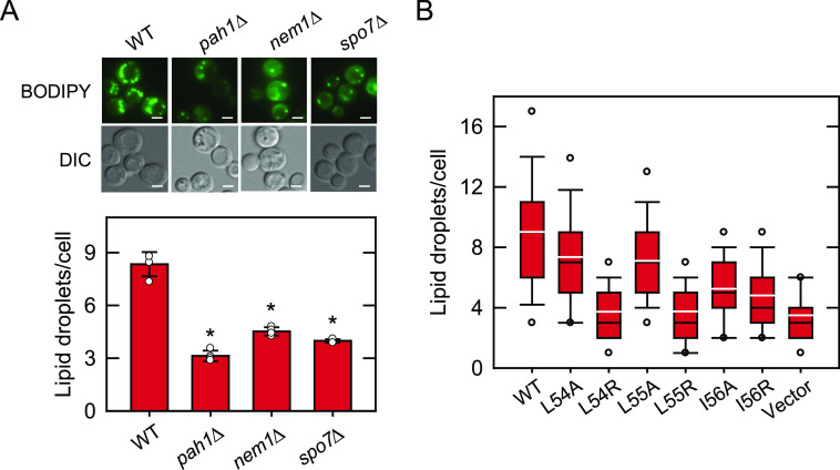
Figure 7.
Lipid droplet formation of cells defective in the Nem1-Spo7/Pah1 phosphatase cascade and of spo7Δ cells expressing Spo7 with LLI mutations. A, WT, pah1Δ, nem1Δ, and spo7Δ cells were grown at 30 °C in SC medium to stationary phase and stained with BODIPY 493/503. B, spo7Δ (GHY68) transformants expressing the indicated SPO7 allele (pGH443 and its derivatives) were grown and stained as in panel A, except that SC-Leu medium was used for plasmid selection. The stained lipid droplets were visualized by fluorescence microscopy, and the number of lipid droplets was counted from ≥4 fields of view (≥200 cells). A, upper, the images shown are representative of multiple fields of view. DIC, differential interference contrast. White bar, 2 μm. A, lower, the data shown are averages from three experiments ± S.D. (error bars). The individual data points are also shown. *, p < 0.05 versus cells expressing the WT control. B, the data are presented by the box plot. The black and white lines are the median and mean values, respectively, and the white circles are the outlier data points of the 5th and 95th percentiles.

