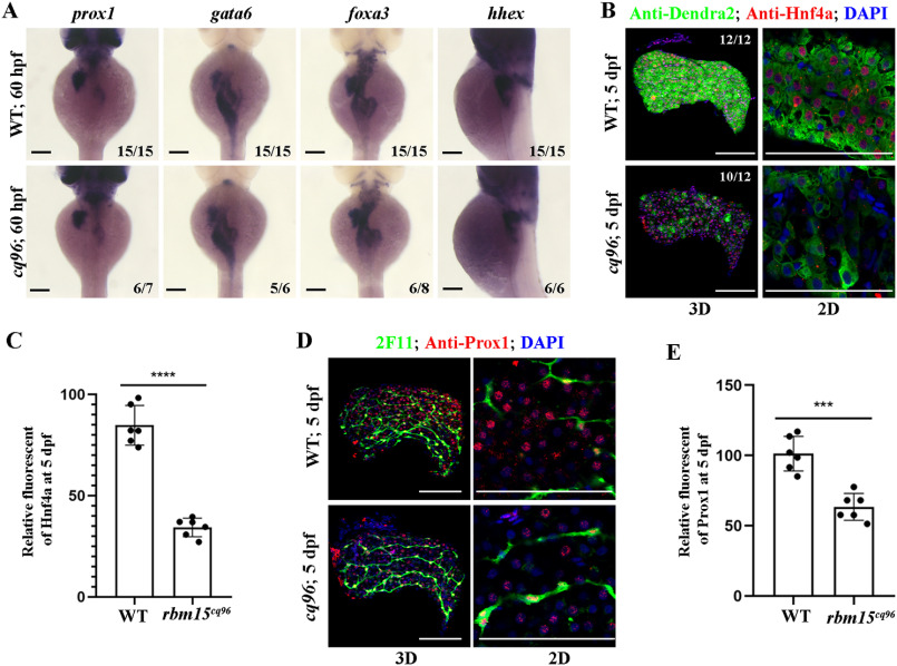
Figure 3.
Hepatoblast specification is normal in the cq96 mutant. A, WISH results of gata6, prox1, foxa3, and hhex at 60 hpf revealing hepatic formation and specification in cq96 and WT. B, confocal images showing the antibody staining of Hnf4a and Dendra2 at 5 dpf in cq96 mutant. C, the quantification of Hnf4a fluorescent intensity in WT and cq96. D, confocal images showing the antibody staining of Prox1 and 2F11 in WT and mutant at 5 dpf. E, the quantification of Prox1 fluorescent intensity in WT and cq96. Numbers indicate the proportion of larvae exhibiting the expression shown. Asterisks indicate statistical significance: ***, p < 0.001; ****, p < 0.0001. Scale bars, 100 μm; error bars, S.D.

