Figure 1.
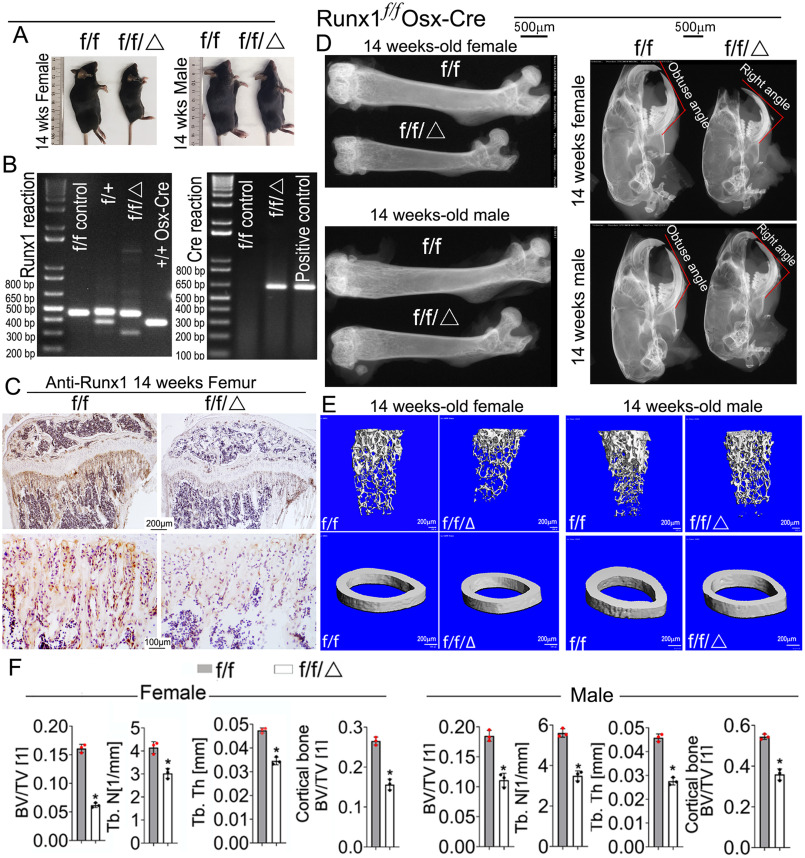
Runx1f/fOsx-Cre mice have decreased bone mineralization and skeletal deformities. A, photographic images of 14-week–old (n = 16) Runx1f/fOsx-Cre (f/f/Δ) mice and WT (f/f) mice. B, PCR was used to determine Runx1 alleles (f/f, f/+, +/+, or deletion) and the presence of Cre. C, IHC staining of 14-week–old Runx1f/fOsx-Cre (f/f/Δ), and WT (f/f) mice tibias using RUNX1 antibody. D, x-ray analysis of 14-week-old male and female femurs and skulls, n = 19. E, μCT scans of femurs from 14-week-old male and female mice, n = 3. F, quantification of E. Results are expressed as mean ± S.D.; *, p < 0.05.

