
Figure 2.
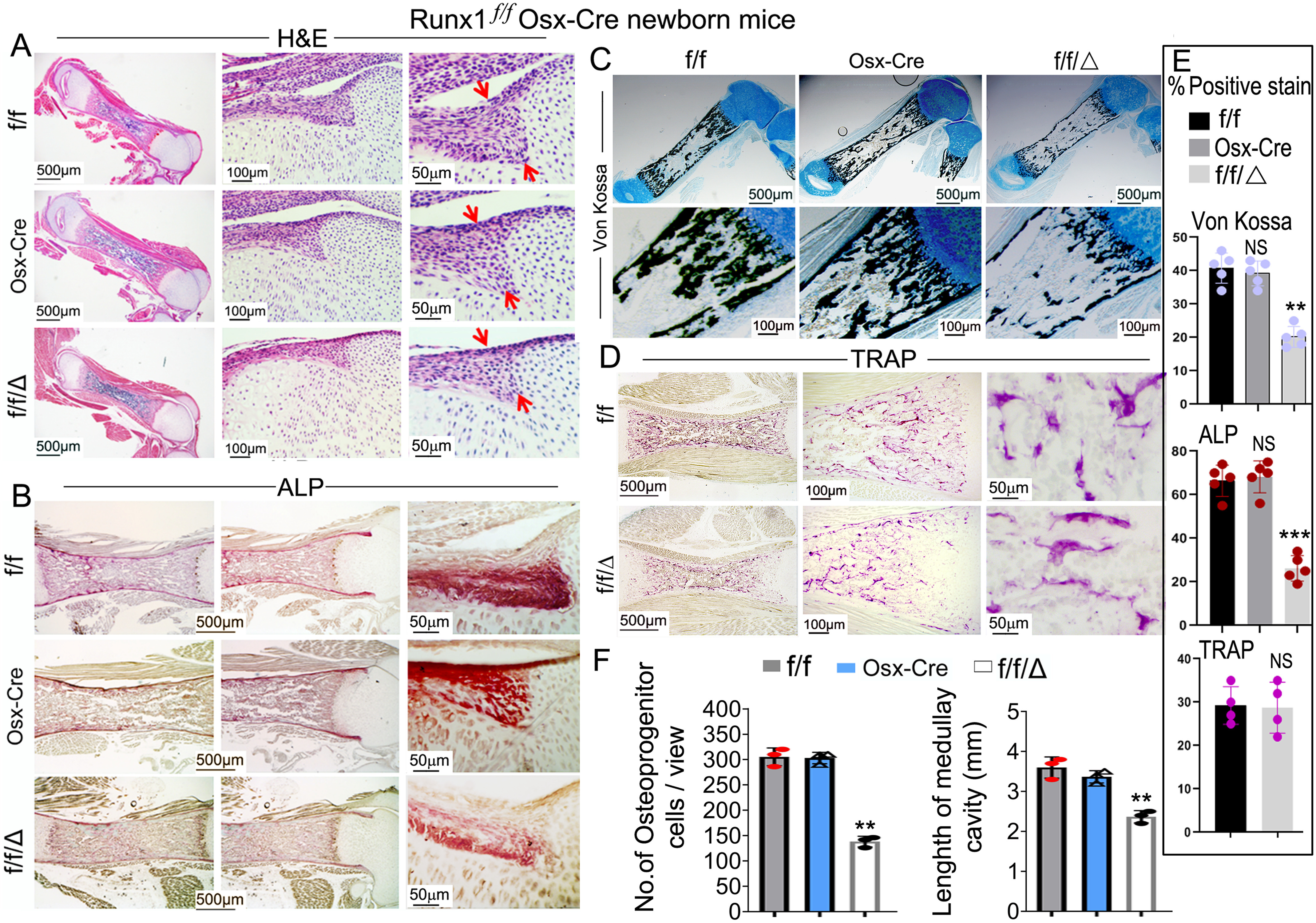
Endochondral bone ossification was significantly impaired and osteoblast numbers were largely decreased in Runx1f/fOsx-Cre newborn mice femurs, but not in Osx-Cre newborn mice femurs compared with WT mice femurs. A–D, newborn Runx1f/fOsx-Cre (f/f/Δ), Osx-Cre and WT (f/f) mice femurs were stained with (A) H&E stain, (B) ALP stain, (C) Von Kossa and Alcian blue stain, and newborn Runx1f/fOsx-Cre (f/f/Δ) and WT (f/f) mice femurs were stained with (D) TRAP stain. E, histomorphometry of WT (f/f), Osx-Cre and Runx1f/fOsx-Cre (f/f/Δ) mice (n = 4) showing ALP stain surface per bone surface (BS) area, Von Kossa stain surface per bone surface area, and TRAP stain surface per bone surface area. F, quantification of cells in the periosteum in A and length of the medullary cavity. Results are expressed as mean ± S.D. NS, not significant; **, p < 0.01; ***, p < 0.001.