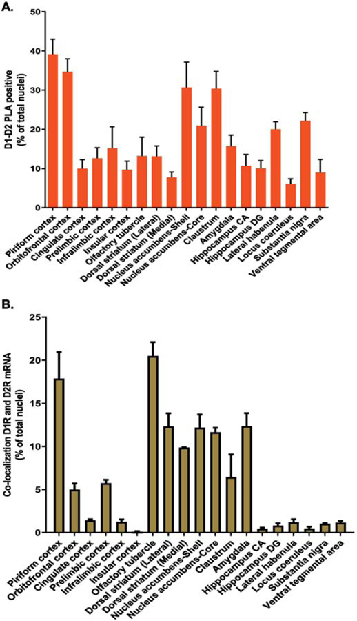
Figure 1. Dopamine D1–D2 receptor heteromer distribution with D1R and D2R mRNA colocalization in rat brain regions.
Serial coronal slices from various rat brain regions were used for the estimation of cells expressing dopamine D1–D2 receptor heteromer using in situ PLA (A), and cells coexpressing D1 and D2 mRNAs using FISH (B). Data resulting from both techniques are presented as the percentage of positive cells per total number of DAPI-labeled nuclei from N=3 male rats.

