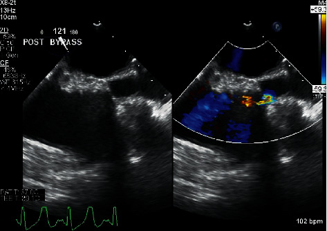
Figure 2.

The image in the left panel shows the initial postbypass two-dimensional presurgical midesophageal bicaval TEE view demonstrating superior vena cava stenosis. The image in the right panel is the same with the addition of color flow Doppler demonstrating flow acceleration across the stenosis.
