
Figure 2.
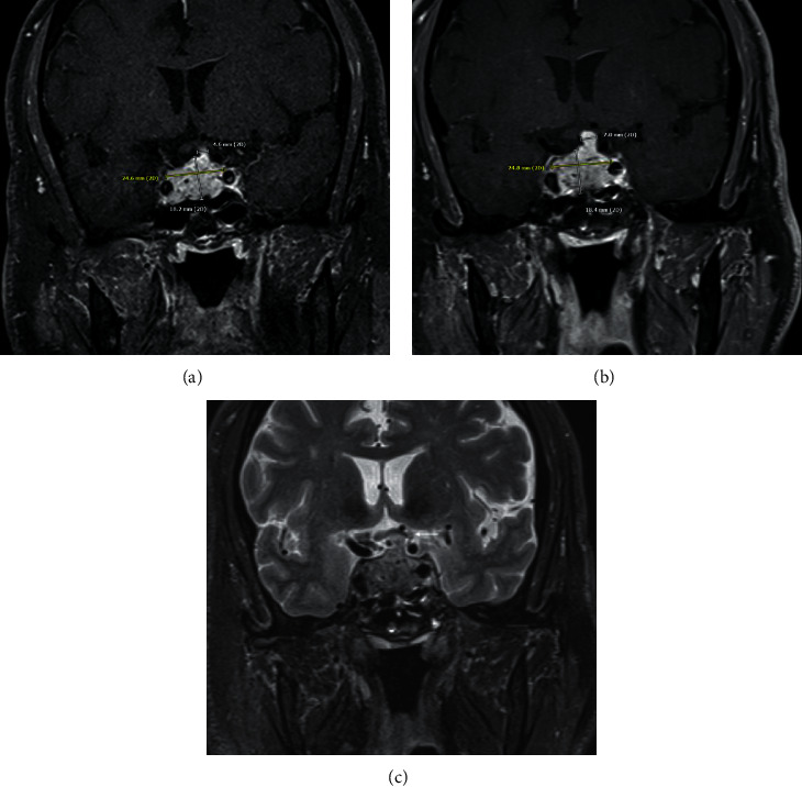
Coronal postcontrast image demonstrates the size of the mass upon initial imaging (a) also including the measurement of the mildly thickened pituitary stalk. At the time of follow-up imaging (b), the mass has slightly increased in size, which is most notable at the pituitary stalk, which now measures 7 mm in thickness, previously 4.6 mm. Also, note the mild increased mass effect on the optic chiasm (thin arrow) at the time of follow-up imaging on the coronal T2 image (c).