

Abstract
Small bowel obstruction from internal hernias is a familiar pathology for the surgeon, with an incidence of 0.5–5.8%. However, pericaecal hernia is a very uncommon type of internal hernia. Diagnosis and early treatment are essential to avoid strangulation and necrosis of the incarcerated small bowel. We report a case of an 84-year-old woman with no previous history of abdominal surgery who came to our hospital having endured 6 hours of abdominal pain and vomiting. Following physical examination and computed tomography, a diagnosis of small bowel obstruction caused by pericaecal hernia was made and emergency surgery was performed. The hernia was successfully reduced with a laparoscopic approach. Although pericaecal hernia is a rare disease, surgeons should bear it in mind as a differential diagnosis in small bowel obstruction.
Keywords: Pericaecal hernia, Paracaecal hernia, Small bowel obstruction, Internal hernia
Introduction
Internal hernias are defined by the protrusion of abdominal viscera through a normal or abnormal peritoneal or mesenteric aperture within the confines of the peritoneal cavity.1 They are an unusual cause of small bowel obstruction with an incidence of 0.5–5.8%.2 Pericaecal hernia is the second most common type of internal hernia. However, few cases have been reported because of its uncommon presentation. Its preoperative diagnosis is difficult to confirm owing to non-specific symptoms and signs. Abdominal computed tomography (CT) can play a key role in the management and diagnosis of patients with intestinal obstruction caused by any type of internal hernia although surgery may be the only option to make an accurate diagnosis and treat this type of hernia. We present a case of small bowel obstruction, provoked by a pericaecal hernia, which was successfully resolved with a laparoscopic approach.
Case history
An 84-year-old woman visited the emergency department after 6 hours of abdominal pain and vomiting. She had a medical history of hypertension, type 2 diabetes mellitus and Alzheimer’s disease, without prior abdominal procedures. On physical examination, her vital signs were stable. She had abdominal distension with tenderness in the right lower abdomen, without rebound or guarding. No abdominal mass or wall hernias were visible or palpable.
Leucocytosis (27.6 x 10⁹/l) with a predominance of neutrophils (95.4%) and elevated C-reactive protein (105mg/l) were observed on laboratory tests. Based on this clinical presentation, abdominal CT was requested, showing dilated small bowel loops, apparently of the distal ileum, with an area of stenosis (transitional zone) trapped in an unusual fossa immediately next to the caecum in the right lower abdomen. There was mesenteric congestion indicating obstruction but there were no signs of ischaemia or infarction (Fig 1). A diagnosis was made of small bowel obstruction due to a probable pericaecal hernia and emergency surgery was performed.
Figure 1. A: Enhanced computed tomography of the abdomen showing cluster of collapsed small bowel (SB) next to the caecum (C): B: Computed tomography showing the caecum displaced inwards by dilated small bowel loop.

A laparoscopic approach revealed 20cm of small bowel loop trapped in a narrow peritoneal fold in the right paracolic gutter. The anterior part of the neck of the hernia was incised by diathermy in order to reduce the loop inside it by exerting traction. In addition, the incision of the hernia sac was enlarged to avoid hernia recurrence. Finally, the colour and the peristalsis of the incarcerated loop were confirmed as correct so intestinal resection was discarded (Video 1 – available online). Postoperative recovery was uneventful and the patient was discharged on day 7.
Video 1.
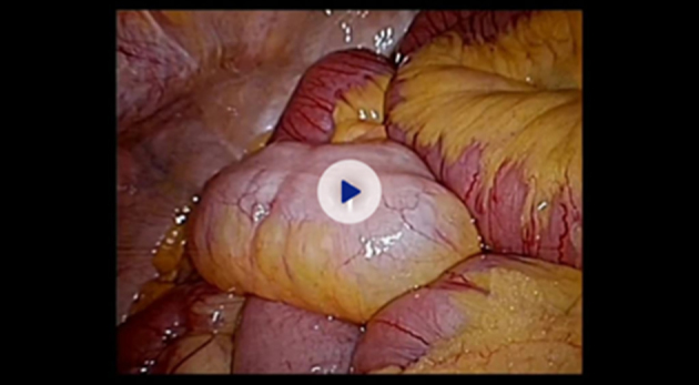
Small bowel obstruction caused by pericaecal hernia resolved with a laparoscopic approach
Discussion
Pericaecal hernia is a type of internal hernia. According to Meyers, internal hernias can be categorised as paraduodenal (53%), pericaecal (13%), foramen of Winslow (8%), transmesenteric and transmesocolic (8%), intersigmoid (6%) and retroanastomotic (5%).1 Pericaecal hernia is relatively rare and accounts for 10–15% of all internal hernias.3 It is produced by a defect in the peritoneum next to the caecum resulting from alterations in the normal process of intestinal rotation during embryonic development. This includes budding, exteriorisation into the umbilicus and subsequent retraction on to the posterior abdominal wall, which usually predisposes the pericaecal fossa to the formation of a number of pockets or recesses.4
Most pericaecal hernias involve an ileal segment protruding through a defective caecal mesentery into the pericaecal fossa and extending towards the right paracolic gutter.1 Meyers divided pericaecal hernias into four subtypes: internal, lateral, retrocaecal recess and unclassifiable.5 Another classification, devised by Endo, divides pericaecal hernias into superior ileocecal, inferior ileocecal, appendicular fossa and retrocaecal recess types.5 The most common site for pericaecal hernias is the retrocaecal fossae, with a reported incidence of 74% among pericaecal hernias.6 In our patient, the hernia orifice and hernia sac were placed laterally to the caecum, meaning the hernia corresponded to the lateral type according to Meyers’ classification and to the retrocaecal type according to Endo’s classification.
Sixteen cases of pericaecal hernia have been reported in the English literature during the last twenty years. Four cases were classified as retrocaecal and two as ileocecal (Table 1). The rest of the reports did not indicate the classification.
Table 1.
Literature review of pericaecal hernia cases published between 2000 and 2019
| Paper | Patient age / sex | Preoperative diagnosis | Classification of internal hernia | Operation performed | Fossa sutured? |
|---|---|---|---|---|---|
| Choh, 2010 | 65 F | Pericaecal hernia | Not available | Laparotomy | Sutured |
| Kabashima, 2010 | 43 F | Small bowel obstruction | Pericaecal | Mini-laparotomy | Not sutured |
| Shibuya, 2010 | 63 M | Small bowel obstruction | Retrocaecal | Laparotomy | Sutured |
| Jang, 2011 | 84 F | Not available | Pericaecal | Laparotomy | Not sutured |
| Nishi, 2011 | 70 F | Internal hernia | Not available | Laparotomy | Not sutured |
| Kleyman, 2013 | 34 M | Small bowel obstruction | Not available | Laparotomy | Not available |
| Kumar, 2015 | 88 F | Small bowel obstruction | Ileocaecal | Laparotomy | Not available |
| Saygin, 2015 | 50 F | Small bowel obstruction | Pericaecal | Laparoscopy | Not available |
| Ogami, 2016 | 92 M | Small bowel obstruction | Retrocaecal | Laparoscopy | Not sutured |
| Sasaki, 2016 | 65 M | Internal hernia | Retrocaecal | Laparoscopy | Sutured |
| Ito, 2017 | 83 M | Retrocaecal hernia | Retrocaecal | Laparotomy | Not sutured |
| Inukai, 2018 | 54 M | Internal hernia | Pericaecal | Laparoscopically assisted | Not sutured |
| Lee, 2018 | 46 M, 55 F | Pericaecal hernia | Pericaecal | No surgery | – |
| Otani, 2018 | 83 F | Paracaecal hernia | Pericaecal | Laparoscopy | Not sutured |
| AlJaberi, 2019 | 16 M | Small bowel obstruction | Ileocaecal | Laparotomy | Not available |
Pericaecal hernias are difficult to diagnose preoperatively because of their uncommon presentation and non-specific clinical manifestations. However, this type of hernia may induce intermittent pain in the right iliac fossa or develop into a palpable mass in this area. As in our case, most patients present with obstruction. The small bowel is the most commonly herniated bowel segment, with a high incidence of mechanical obstruction and fast progression to strangulation.1 Despite the lack of specific symptoms, abdominal CT is an important tool for diagnosis. Plain radiography and barium enhanced studies might demonstrate signs of bowel obstruction or indirect signs of internal hernia. Nevertheless, CT appears to be highly accurate in the diagnosis of internal hernias. Conversely, visceral internal herniation is often only discovered with laparotomy. In our literature review, CT was able to aid in the diagnosis of internal hernia or pericaecal hernia in seven cases.
Treatment for small bowel obstruction caused by a pericaecal hernia is mainly in the form of surgery. A laparoscopic approach has been found to be useful for the diagnosis and treatment of internal hernias. Furthermore, a systematic review comparing laparotomy and laparoscopy for obstruction showed that laparoscopy results in lower morbidity, shorter postoperative hospital stay and faster return of bowel function.6 On the other hand, laparoscopy for intestinal obstruction is a demanding procedure that should be performed by an experienced surgeon. In our case, surgical treatment was laparoscopic and there were no complications during or after surgery. Six of these sixteen cases in our literature review were resolved through laparoscopic surgery (Table 1). Regarding the hernia orifice, opening or closing it are both feasible options. While we decided to open the hernia neck, enlarging the pericaecal fossa to avoid recurrence, some authors have reported closing the defect with satisfactory results.2
Conclusions
Although pericaecal hernia is a very uncommon cause of intestinal obstruction, this pathology should be considered as a differential diagnosis because of its rapid progression to strangulation and necrosis. Laparoscopic treatment appears to be a viable technique for this condition.
References
- 1.Martin LC, Merkle EM, Thompson WM. Review of internal hernias: radiographic and clinical findings. Am J Roentgenol 2006; : 703–717. [DOI] [PubMed] [Google Scholar]
- 2.Choh NA, Rasheed M, Jehangir M. The computed tomography diagnosis of paracecal hernia. Hernia 2010; : 527–529. [DOI] [PubMed] [Google Scholar]
- 3.Lee JE, Choi SY, Lee MH et al. Pericecal herniation of sigmoid colon diagnosed by computed tomography. Medicine 2018; : e11336. [DOI] [PMC free article] [PubMed] [Google Scholar]
- 4.Jang EJ, Cho SH, Kim DD. A case of small bowel obstruction due to a paracecal hernia. J Korean Soc Coloproctol 2011; : 41–43. [DOI] [PMC free article] [PubMed] [Google Scholar]
- 5.Otani H, Makihara S. Laparoscopic surgery for small bowel obstruction due to paracecal hernia. Acta Med Okayama 2018; : 81–84. [DOI] [PubMed] [Google Scholar]
- 6.Ogami T, Honjo H, Kusanagi H. Pericecal hernia manifesting as a small bowel obstruction successfully treated with laparoscopic surgery. J Surg Case Reports 2016; : 1–4. [DOI] [PMC free article] [PubMed] [Google Scholar]