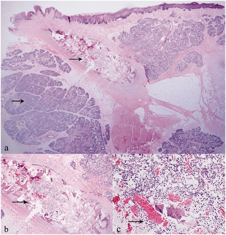
Figure 3.
Histopathological images of lingual hamartoma: (a) salivary gland hyperplasia and localized fibrous tissue with ossification (arrow) (HE, 12.5×); (b) ossification with bone marrow-like tissue formation (arrow) (HE, 40×); (c) bone marrow cells and dilated capillaries (arrow) (HE, 200×).
Abbreviation: HE, hematoxylin and eosin.

