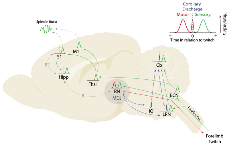
Figure 1.
The neural causes and consequences of myoclonic twitching in sleeping infant rats. Inset: Illustration of neural activity surrounding a twitch: preceding activity indicative of motor outflow (red), following activity indicative of sensory feedback (i.e., reafference; green), and nearly simultaneous activity indicative of corollary discharge (blue). These icons are used in the main figure below, which is based largely on studies using P8 rats. Main figure: The production of a forelimb twitch begins in midbrain structures, including the red nucleus (RN) and surrounding areas in the mesodiencephalic junction (MDJ). When a forelimb twitch is produced, reafferent signals flow directly to the spinal cord, the external cuneate nucleus (ECN), and the RN. From the ECN, reafferent signals flow to the deep cerebellar nuclei and cerebellar cortex (Cb) as well as the thalamus, arriving next in primary somatosensory (S1) and primary motor (M1) cortex. From S1, reafference flows via the entorhinal cortex to the hippocampus. Coincident with spiking activity in S1 and M1, spindle bursts are detected in the local field potential. Corollary discharges emanating from neurons in and around the RN project separately to the inferior olive (IO) and lateral reticular nucleus (LRN), before projecting to the cerebellum via climbing and mossy fibers, respectively. Finally, the occurrence of twitch-triggered bursts of coherent rhythmic activity between RN and hippocampus in the theta band (θ; 4–7 Hz) and between S1 and hippocampus in the beta2 band (β2; 20–30 Hz) is illustrated. Background image is a sagittal section of an infant rat brain. See text for discussion and citations.

