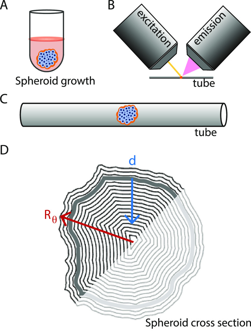
Figure 2.
Overview of the experimental setup. (A) Illustration of gravitation-assisted tumor spheroid growth. (B) Sketch of a spheroid mounted for imaging in an Airy beam light-sheet microscope, with one objective for the (yellow) light sheet (excitation) and one for collection of the emitted light (emission). (C) Close-up of a spheroid mounted in a tube for imaging. (D) Example of a spheroid’s cross-section mask, where the radial vector from the origin goes to the mask’s boundary; Rθ varies in length with respect to the polar angle θ. The mask boundary was slightly larger than the spheroid’s surface (gray contour) and the blue vector indicates the tumor depth, d. We restricted our analysis to the half-spheroid closest to the light-sheet objective (excitation).

