
Extended Data Figure 10. Rreb1−/− mouse embryo chimeras exhibit defects in early development.
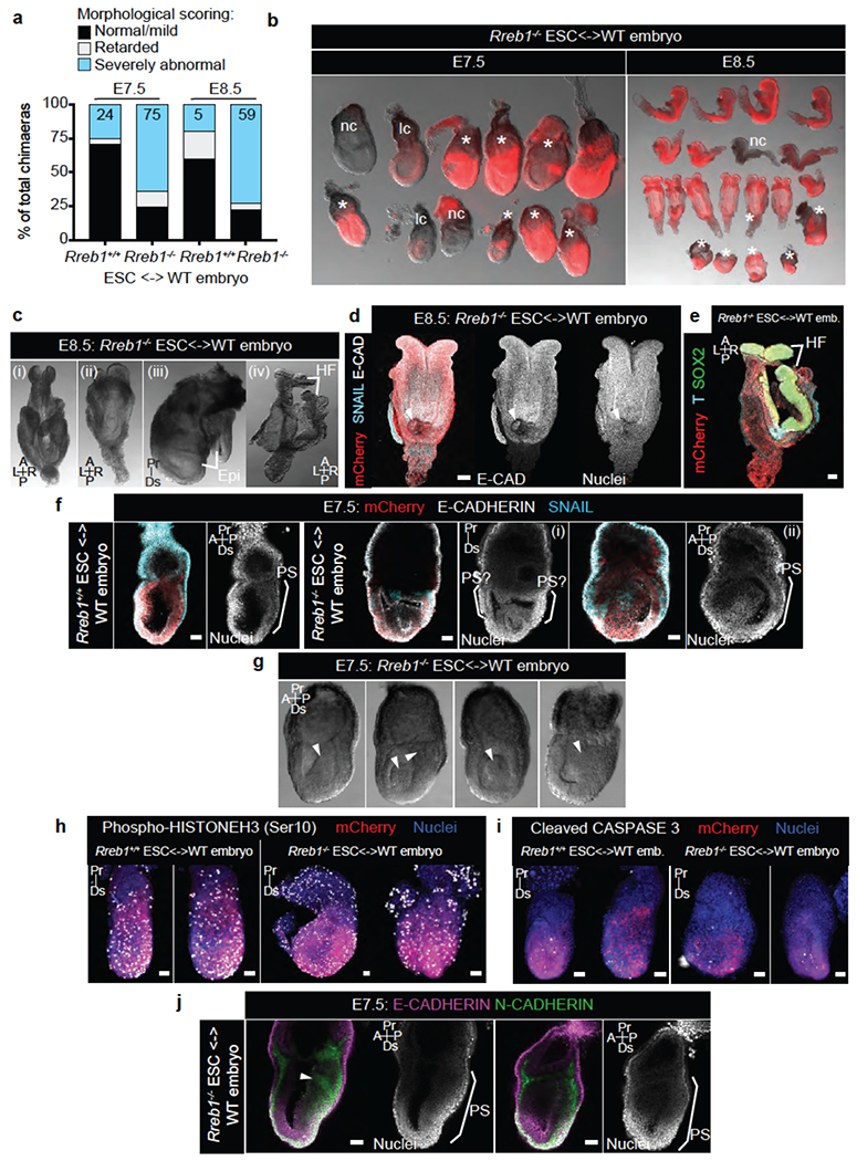
(a) E7.5 and E8.5 chimeric embryos containing WT ESCs or Rreb1−/− ESCs were scored, based on gross morphology, as normal/mild defects, developmentally retarded or severely abnormal. At E7.5, a fraction of Rreb1+/+ ESC embryos displayed small clumps of cells in the amniotic cavity, possibly an artifact from the microinjection, and hence were scored as abnormal. Rreb1+/+ data is compiled from 4 distinct KO clones, (b) Images showing brightfield morphology and mCherry fluorescence (marking descendants of injected ESCs) in representative litters of Rreb1+/+ ESC-containing chimeric embryos dissected at E7.5 and E8.5. nc, non-chimeric; lc, low chimerism. Asterisks mark morphologically abnormal/developmentally retarded embryos, (c) Brightfield images of morphologically abnormal Rreb1−/− ESC-containing chimeric E8.5 embryos. Embryos exhibited abnormal headfold development including disproportionate headfolds (i), asymmetric headfolds (ii). Axis duplication was also observed (iii) and (iv). To note, the embryo in panel (iii) is also developmentally retarded. (d)(e) Confocal maximum intensity projections of wholemount immunostained E8.5 Rreb1−/− ESC-containing chimeric embryos. Panel (d), an embryo with an ectopic somite-like structure (arrowhead). Panel (e), the embryo in (c)(iv) with axis duplication of the headfolds. (f) Sagittal confocal optical sections of wholemount immunostained chimeric E7.5 embryos. Embryos shown in (f)(i)-(ii) have multiple cavities and multiple expression sites of SNAIL hence anterior-posterior axis orientation is not possible, (g) Brightfield images of morphologically abnormal Rreb1−/− ESC-containing chimeric E7.5 embryos. Embryos frequently had protrusions into the cavity and thickening of the posterior epiblast, marked by arrowheads. (h)(i) Confocal maximum intensity projections of chimeric embryos after wholemount immunostaining for phospho-Histone H3 (h), labeling mitotic cells, and cleaved Caspase 3 (i) labeling apoptotic cells. Brackets demarcate the primitive streak, (j) Sagittal confocal optical sections of chimeric E7.5 embryos after wholemount immunostaining for E-cadherin and N-cadherin. Arrowhead, aberrant N-cadherin expression. HF, headfold; PS, primitive streak; A, anterior; P, posterior; Pr, proximal; Ds, distal; L, left; R, right. Scale bars, 50 μm. (b-j) Images are representative of two independent experiments.