Figure 5.

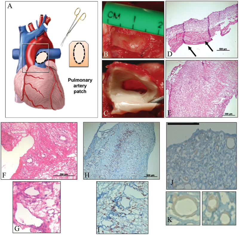
In vivo experiments assessed the functionality of hybrid scaffolds under physiological pressure and stress. A) Schematic of the hybrid patch sized, cut, and placed as a patch on sheep pulmonary artery. Actual images from surgical patch implant, both with sutures, prior to explant B) and post-explant of patch with native tissue attached C). Images D) and E) portray standard H&E stains of the hybrid scaffold with tissue formation, post-explant, in cross-sectional D) and surface E) orientations. F,G) Magnifications of the H&E stains show presence of cells and sites of potential lumen formation. H,I) Hybrid scaffolds were also stained for α-SMA to confirm cell integrity and motility. The majority of cells were within the interior of the scaffold. J,K) Stains for CD45 were performed to confirm the presence of various cell types, if any, but the stain was inconclusive; magnifications K) suggest possible sites where cells are beginning to line preliminary lumen.
