
Figure 3.
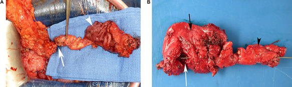
Gross examination of a surgical specimen. (A) Perioperative view of the complete resected tissue, showing the normal gastric mucosa (white arrowhead) and the duplicated head of pancreas (white arrow) before cutting the duct. (B) View of the complete specimen, including the gastric duplication (opened, white arrow), the gastric wall (side of the serosa, black arrow) and the duplicated head of pancreas (black arrowhead) containing a duplicated duct catheterized from the cut on pancreas to the gastric duplication with a metal probe.