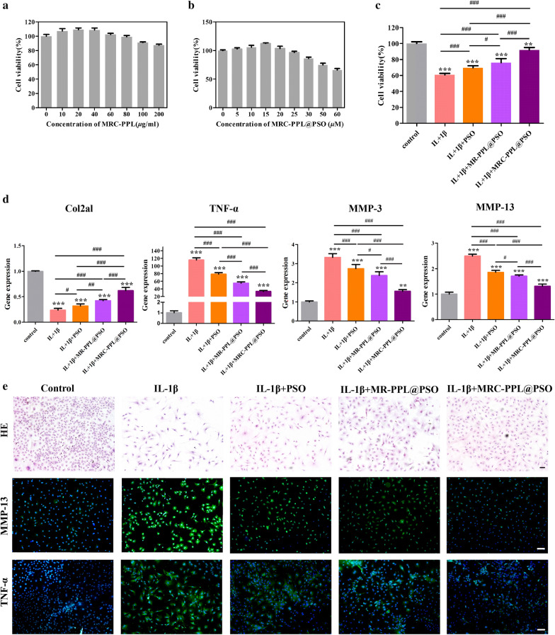
Fig. 3.
In vitro study on chondrocytes isolated from C57BL6/J mice. (a and b) Cell viability after treatment with MRC-PPL or MRC-PPL@PSO. (c) Cell viability after various treatments to IL-1β-stimulated chondrocytes. (d) Relative mRNA levels of Col2a1, TNF-α, MMP-3 and MMP-13 on IL-1β-stimulated chondrocytes with various treatments. (e) HE staining and immunofluorescence images. The nuclei were counterstained with DAPI (blue), and MMP-13 or TNF-α positive staining was stained with FITC (green). Scales bar: 400 μm. Each data point represents mean ± s.d (n = 3). *, # indicate p < 0.05; **, ## indicate p < 0.01; ***, ### indicate p < 0.001

