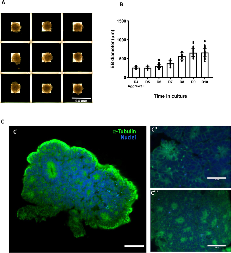
Fig. 18.
Embryoid body generation and size over time. A) Four day-old embryoid bodies (EB) generated using microwells from Aggrewell. B) Relative growth of the embryoid bodies over time. C) Light sheet microscope images of day 7 embryoid body showing the formation of organized neural rosettes. C” and C” show close up to the neural rosettes. Data are mean ± s.d. Scale bars: (A) 0.5 mm, (C’) 100 lm, (C”) 50 lm, (C”’) 100 μm.

