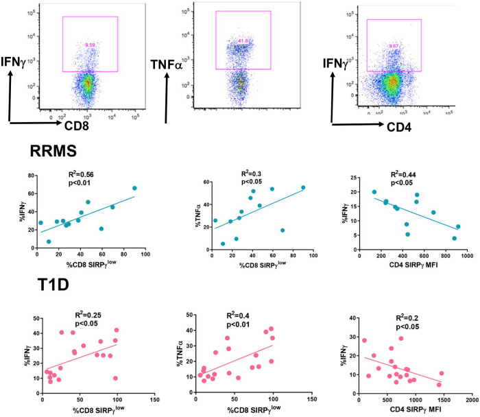
Fig 4. SIRPγlow T-cells positively correlate with proinflammatory molecules in subjects with autoimmunity.
PBMCs from RRMS and T1D patients were activated with PMA/INO in the presence of golgi plug for 6 hrs. Representative flow plots for IFNγ and TNFα staining on T-cells are shown. Following stimulation, cells were stained extracellularly with fluorescently tagged anti-CD3, CD4, CD8, and SIRPγ, followed by intracellular staining with anti-IFNγ, and anti-TNFα. Data was correlated with Pearson’s test and p<0.05 was considered significant.

