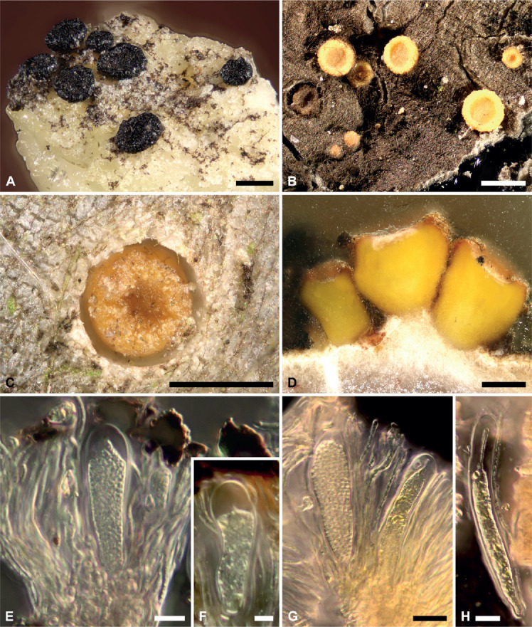
Fig. 1.

Light micrographs of Sarea difformis and S. resinae. A. Ascomata of S. difformis and B. S. resinae; C. Young ascoma of S. resinae arising on a fresh resin flow; D. Cross-section of S. resinae showing hyphal growth into the liquid resin; E. Ascus and paraphyses of S. difformis; F. Young ascus of S. difformis; G. Asci and paraphyses of S. resinae; H. Young ascus of S. resinae. Scale bars: 1 mm (A, B), 500 μm (C, D), 10 μm (E, G), 5 μm (F, H).
