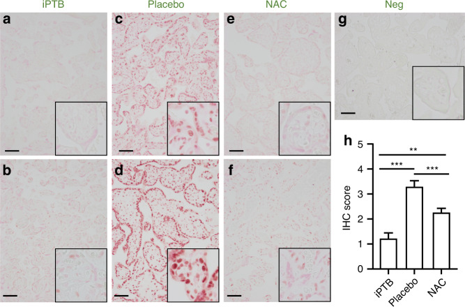
Fig. 2. Immunohistochemistry of placental villous tissue for histone deacetylase-2.
Representative micrographs of immunohistochemical staining for histone deacetylase-2 (HDAC2) in placental villous tissue from women with idiopathic preterm birth (iPTB, a, b) absent Triple I or PTB in the context of Triple I who were enrolled in the trial and received either placebo (c, d) or N-acetylcysteine infusion (e, f). Vector NovaRed was used as peroxidase substrate and tissues were imaged and scored blindly for staining intensity without counterstaining. Negative slides (g) were exposed to nonimmune serum. Higher magnification inserts are shown in each lower right corner. All tissues available from the subjects in the trial were analyzed (NAC: n = 32; placebo: n = 33). **P < 0.01; ***p < 0.001. The cases presented in a, c, e, and g were delivered at 28 weeks of gestation. The cases illustrated in b, d, and f were delivered at 32 weeks of gestation. The scale bar (50 μm) denotes the magnification for the panels, which were photographed at ×200. Insets were imaged at ×600 magnification.

