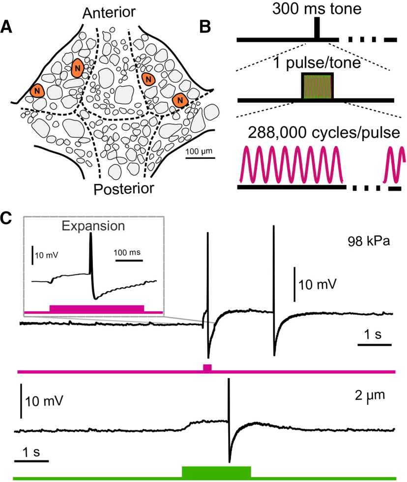
Figure 5.
US application and electrode displacement yield similar results when a different neuron (N cell) and different pulse parameters are used. A, Schematic of ventral surface of a single leech ganglion with N neurons marked. B, US parameters applied to N cells. We applied one tone (300-ms duration) of continuous (vs pulsed) US per trial. C, Representative intracellular traces of N cell voltage during a trial of US application (upper, pink) and electrode displacement (lower, green). When upper trace is expanded (inset), the waveform closely resembles that observed in the electrode displacement paradigm. The difference in the duration of the US-induced depolarization can be attributed to the difference in stimulus duration.

