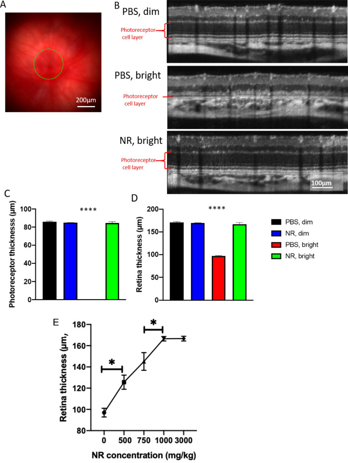
Figure 4.
NR treatment preserves photoreceptor layer thickness and total retinal thickness as assessed in vivo following LIRD. (A) Representative fundus image. (B) Representative OCT images from each group. The OCT image is a circular scan about 100 µm from the optic nerve head. Photoreceptor thickness (C) and retinal thickness (D) from BALB/c mice 1 week after retinal degeneration induction. Mice treated with PBS and exposed to 3000-lux light for 4 hours (red bar) exhibited great losses in thickness of the photoreceptor and retina layers, whereas induced mice treated with NR (green bar) exhibited statistically significant preservation of layer thickness. (E) Treating with increasing concentrations of NR up to 1000 mg/kg results in significantly increasing retina thicknesses in mice exposed to 3000-lux light for 4 hours. *P < 0.05 between two adjacent groups; ****P < 0.0001 versus all other group means; one-way ANOVA with Newman-Keuls multiple comparisons post hoc test. N = 3–9 mice per group. Error bars represent SEM. Size marker = 200 µm or 100 µm.

