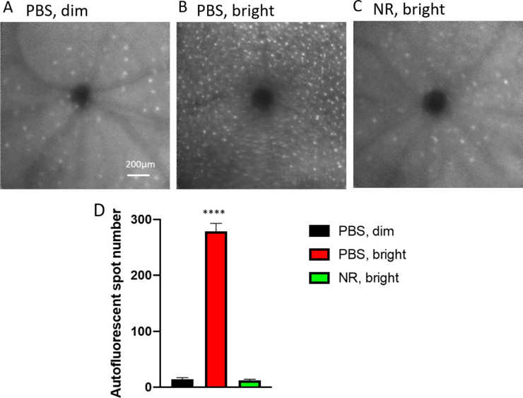
Figure 7.
NR treatment prevented subretinal autofluorescence observed in vivo in LIRD mice. (A–C) Representative morphology images from each group at the level of the photoreceptor-RPE interface. In vivo Spectralis HRA+OCT images (with autofluorescence detection) were taken 1 week after induction of degeneration. (D) Autofluorescent spots were counted across the fundus image field. Few were detected in uninduced mice (black). Toxic light-exposed mice treated with NR exhibited significantly fewer autofluorescent spots compared with the PBS treated group. ****P < 0.0001 one-way ANOVA with Newman-Keuls multiple comparisons post hoc test. N = 3–6 mice per group. Error bars represent SEM. Size marker = 200 µm.

